
Originally published in February 2013 and comprehensively updated and revised in July 2026.
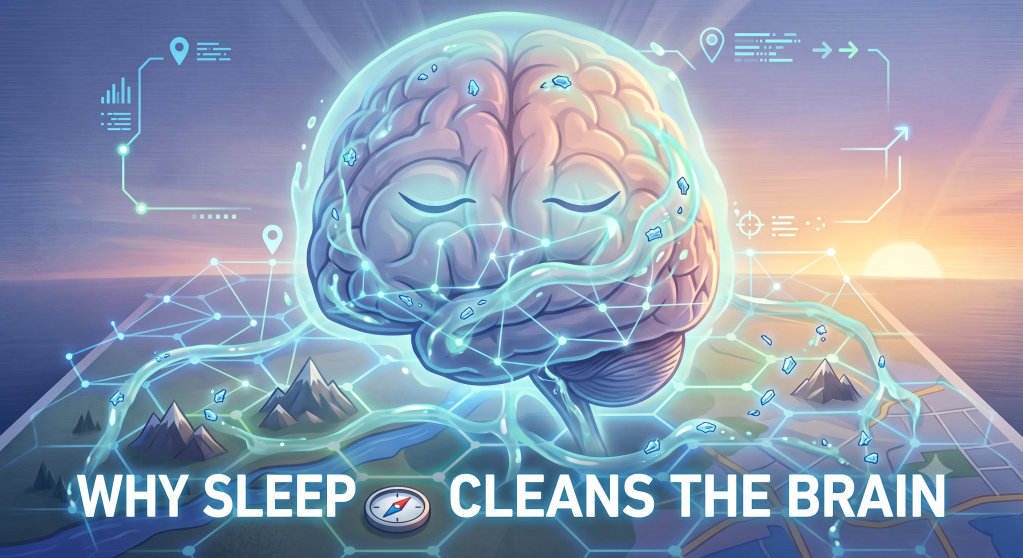
How the discovery of the brain’s glymphatic system revealed a possible mechanism behind sleep’s restorative power and transformed research on brain health and Alzheimer’s disease.
In the early months of 2013, sleep still had an image problem. American culture had spent the better part of a decade celebrating the idea that rest was optional for anyone ambitious enough to skip it. Certain corporate cultures celebrated employees who presented extreme sleep deprivation as evidence of ambition and commitment. Silicon Valley executives spoke openly about sleep as a design flaw to be engineered around, not a biological necessity to be respected. The prevailing scientific answer to the question “why do we sleep at all” was still frustratingly incomplete. Researchers understood that sleep supported memory, metabolism, and physiological restoration, but no one could point to a single, concrete, physical process that explained why a brain deprived of sleep began to fail so quickly and so completely.
Then, in the fall of 2013, a paper published in the journal Science offered something the field had been missing for decades: a plumbing system. A team led by neuroscientist Maiken Nedergaard at the University of Rochester described a previously unrecognized network of channels that flushes waste out of the brain, and that network turns out to work far harder while we sleep than while we are awake. Researchers already understood many of sleep’s functions, including memory consolidation, metabolic regulation, and emotional processing, but no one had identified a single physical maintenance process that could explain why prolonged sleep deprivation was so damaging to the brain. The researchers called this newly mapped system the glymphatic system, a name that blends “glial,” referring to the brain’s support cells, with “lymphatic,” referring to the body’s waste-clearing plumbing everywhere else. The discovery reframed sleep not as a passive shutdown but as an active maintenance shift, something closer to a nightly cleaning crew than a simple power-down.
This article traces how that idea emerged, what the glymphatic system actually does, and why a discovery about fluid dynamics in the brains of mice ended up reshaping how scientists, and eventually the public, think about insomnia, aging, dementia, and the basic human need to close one’s eyes for roughly a third of every day.
A Body That Cleans Itself, Except in One Place
To understand why the glymphatic discovery mattered, it helps to understand a puzzle that had quietly bothered physiologists for a long time. Every organ in the human body produces metabolic waste. Muscles generate byproducts during exercise. Kidneys filter toxins from the blood. Nearly every tissue in the body relies on the lymphatic system, a network of vessels that runs alongside the bloodstream and drains away excess fluid, proteins, and cellular debris.
The brain is different. It has no lymphatic vessels of its own. For most of modern medical history, this absence was treated as a minor anatomical footnote rather than a mystery worth solving, largely because the brain is metabolically expensive enough already, burning roughly a fifth of the body’s energy despite making up about two percent of its weight. A high-energy organ produces a great deal of waste, including a protein fragment called amyloid-beta, the same molecule that clumps into the plaques associated with Alzheimer’s disease. If the brain had no dedicated drainage system, where was all of that waste actually going?
For decades, the working assumption was that waste simply diffused out slowly, molecule by molecule, the way a drop of ink might eventually spread through a still glass of water. Diffusion works, but it is slow, and the brain’s waste products, left in place too long, are neurotoxic. Something faster had to be happening. Nedergaard’s team set out to find it using two-photon microscopy, a technique that allows researchers to watch fluid movement in the living brains of mice in real time.
Finding the Brain’s Hidden Plumbing
What the researchers found was a system of channels running along the outside of blood vessels throughout the brain, formed by the end-feet of star-shaped support cells called astrocytes. These channels create a kind of parallel plumbing network, distinct from blood vessels themselves, through which cerebrospinal fluid, the clear liquid that cushions the brain and spinal cord, is pumped inward along the arteries and then pushed back outward along the veins, sweeping through brain tissue in between. Along the way, that fluid picks up metabolic waste, including amyloid-beta, and transports it toward clearance pathways outside the brain.
The team published their initial description of this pathway in 2012, and it drew some attention within the neuroscience community. It was the follow-up study a year later, published in Science in October 2013, that turned the discovery into a genuine sensation. In that paper, lead author Lucy Xie and her colleagues in Nedergaard’s lab measured how quickly the glymphatic system cleared a tracer dye from the brains of sleeping mice compared to brains of mice that were awake. <cite index=”3-1″>The study found that the glymphatic system is highly active during sleep, clearing away toxins associated with Alzheimer’s disease and other neurological disorders.</cite> Just as strikingly, the researchers observed that brain cells physically shrink during sleep, opening up the extracellular space between them by roughly sixty percent in the mouse studies, which allows fluid to flow through brain tissue far more freely than it can when those cells are swollen to their waking size.
That second finding is what made the story so compelling to a general audience. It was not simply that some vague clearing process sped up overnight. The brain appeared to be reorganizing its own architecture, shrinking its cells like a night-shift crew moving furniture aside to mop the floor underneath, in order to make room for a clearance process that could not occur as efficiently under waking conditions.
Why the Brain Can’t Do Both Jobs at Once
The deeper implication of the 2013 paper was a possible answer to one of neuroscience’s oldest open questions: why does sleep exist at all, given how vulnerable it makes an animal to predators and how much time it consumes across an entire lifespan? Nedergaard’s team proposed that the brain faces a genuine energy and space trade-off. Processing information, forming memories, and coordinating the trillions of signals involved in conscious thought all require densely packed, active brain tissue. Efficient clearance may depend on changes in tissue structure that allow fluid to move more freely through the brain. A brain, in other words, may face biological trade-offs between intensive information processing and intensive maintenance. Sleep, under this framework, is what happens when the brain temporarily sets aside the demands of active cognition in order to run its maintenance cycle instead.
It is worth being precise about what kind of claim this is. The idea that sleep exists specifically because the brain needs dedicated, undistracted time to clear waste is a hypothesis, a promising and influential one, but a hypothesis nonetheless. It is not a settled fact in the way that, say, the existence of cerebrospinal fluid itself is a settled fact. Sleep almost certainly serves several distinct biological functions simultaneously, including memory consolidation, hormonal regulation, and emotional processing, and researchers have not established that waste clearance is the single dominant reason sleep evolved. What the 2013 paper did establish, with reasonably strong physical evidence, is that glymphatic clearance really does increase substantially during sleep in mice, and that this increase is tied to measurable changes in brain cell volume and fluid flow.
From Mouse Brains to Human Anxiety About Sleep
The glymphatic discovery arrived at a cultural moment that was primed to receive it. Wearable sleep trackers were just beginning to enter the consumer market, and public health messaging around sleep deprivation was intensifying alongside a growing body of research linking short sleep to obesity, cardiovascular disease, and impaired cognitive performance. Into that environment came a scientific finding that offered something rare: a vivid, almost mechanical image of what insufficient sleep might be doing inside a person’s skull, night after night, as waste products that should have been cleared away instead lingered and accumulated.
This is where the science press, quite understandably, ran ahead of the data. Headlines describing sleep as the brain’s “dishwasher” or “power wash” appeared throughout 2013 and 2014, and Nedergaard herself has used similarly vivid metaphors in interviews. Metaphors like these are genuinely useful for explaining a complex fluid dynamics process to a general audience, but they also invite a subtle overreach: the assumption that a single bad night of sleep leaves measurable toxic buildup, or that any amount of sleep loss meaningfully raises a person’s dementia risk. Neither claim was established by the original mouse studies, which examined the difference between fully awake and fully asleep states rather than gradations of partial sleep loss in humans.
What the glymphatic research did responsibly support was a plausible biological mechanism connecting chronic, severe sleep disruption to the kinds of protein accumulation seen in neurodegenerative disease. That is meaningfully different from a mechanism connecting occasional short nights to measurable brain damage, and the distinction matters for anyone trying to think clearly about their own sleep habits rather than simply feeling anxious about them.
The Amyloid Connection and Alzheimer’s Research
The link to Alzheimer’s disease deserves its own closer look, because it became the most consequential thread to emerge from the glymphatic discovery. Amyloid-beta plaques are one of the defining physical features found in the brains of people with Alzheimer’s disease, though researchers still debate exactly how central a causal role these plaques play in the disease process itself, as opposed to being a downstream marker of some other underlying problem. Regardless of that ongoing debate, if the glymphatic system is one of the brain’s primary routes for clearing amyloid-beta, and if that system runs mainly during sleep, then chronic poor sleep could plausibly interfere with the brain’s ability to clear a protein already implicated in dementia.
Subsequent research through the 2010s explored this connection using human subjects, primarily through indirect measures like PET imaging and cerebrospinal fluid sampling, since directly visualizing glymphatic flow in a living human brain the way researchers can in mice remains technically difficult. Several studies found associations between poor sleep quality, particularly reduced deep slow-wave sleep, and higher levels of amyloid-beta markers in cerebrospinal fluid and brain imaging. These findings are correlational. They show that poor sleep and amyloid accumulation tend to occur together, not that poor sleep definitively causes amyloid accumulation, and researchers in the field have generally been careful to say so, even as public discussion of the findings has sometimes flattened that nuance into more confident-sounding claims.
Refining the Model: What Changed Between 2013 and 2026
Science rarely stands still, and the glymphatic field is a useful reminder of how an influential discovery gets tested, refined, and occasionally challenged over the following decade rather than simply being accepted wholesale. Since 2013, researchers have extended the original findings in several directions while also surfacing genuine methodological disagreements that are worth understanding honestly rather than glossing over.
On the confirming side, later work from Nedergaard’s group and others has shown that glymphatic function appears to decline with age, and separately after traumatic brain injury, both of which offer plausible mechanisms connecting head trauma and aging to elevated dementia risk. Researchers have also identified that certain sleep positions, side-sleeping in particular, may promote more efficient glymphatic clearance in animal studies, along with evidence that specific stages of deep sleep matter more than total sleep time alone.
At the same time, the field has not been without genuine scientific disagreement, and a responsible account of this topic should say so plainly. Some research groups using different tracer methods and imaging techniques have reported findings that complicate the original sleep-clearance story, including at least one study suggesting that clearance to certain lymphatic vessels can actually be faster during wakefulness in some measurement conditions, and more recent work using tracers injected directly into brain tissue rather than cerebrospinal fluid that has produced results conflicting with the original 2013 findings. These disagreements have become sharp enough that some researchers now describe parts of the glymphatic hypothesis as actively contested rather than settled, while Nedergaard and colleagues who favor the original model have pushed back hard on the methodology behind some of the newer, conflicting studies. This is not evidence that the glymphatic system does not exist or that sleep is unrelated to brain waste clearance; the existence of the paravascular fluid channels themselves is well established. It is evidence that the precise relationship between sleep, wakefulness, and the rate of clearance is more complicated and less fully resolved than the simplest version of the “sleep is a dishwasher” story suggests, and that the field is still actively working out the details.
This kind of back-and-forth is normal, healthy science, not a sign that the original discovery was wrong. It does mean that anyone writing or reading about the glymphatic system in 2026 should hold the sleep-clearance connection as a well-supported but still-evolving model rather than a fully closed case.
What This Meant for How People Thought About Rest
Beyond the laboratory, the cultural effect of the glymphatic discovery was significant, even if it unfolded gradually rather than overnight. Through the mid and late 2010s, public health messaging increasingly framed sleep not as downtime but as a biologically active process with its own distinct purpose, comparable to digestion or wound healing. This reframing gave people a more concrete, almost visual way of understanding why chronic sleep deprivation felt so distinctly bad, beyond simple tiredness. It was not just that a sleep-deprived brain was running on empty. It was that a sleep-deprived brain might be running dirty, with waste products that may not be cleared as efficiently because the processes involved in clearance have been disrupted.
Workplace and cultural attitudes shifted alongside the science, though slowly and unevenly. The instinct to treat sleep as a weakness to be overcome did not disappear, but it did face growing pushback from public health authorities, physicians, and eventually corporate wellness programs that began explicitly citing brain-clearance research when making the case for adequate rest. Sleep tracking technology matured considerably over this period as well, with consumer devices increasingly marketing themselves around deep sleep and slow-wave sleep specifically, the stages most closely tied to glymphatic activity, rather than simply tracking total hours slept.
Conclusion: A Maintenance Cycle Written Into Biology
The discovery of the glymphatic system gave neuroscience something it had lacked for a long time: a physical explanation for part of the question of why sleep is not optional. Sleep, according to this research, is not simply the absence of wakefulness. It appears to be an active state during which the brain reorganizes its own cellular architecture to permit a flushing process that clears away the metabolic byproducts of a day’s worth of thought.
More than a decade on, the core finding has held up reasonably well even as researchers have refined, complicated, and in some corners contested the details. The broad picture, that fluid movement through brain tissue changes meaningfully between sleep and wakefulness, and that this movement matters for long-term brain health, remains one of the more consequential ideas to emerge from sleep science in the last fifteen years. Its full implications for preventing or slowing neurodegenerative disease are still being worked out, and will likely take another decade or more of careful human research to establish with real confidence.
What the discovery ultimately offered was not a simple health hack or a clean story about brain hygiene, but a genuine shift in how scientists think about the basic architecture of rest. A brain that shrinks its own cells every night to make room for a cleaning cycle is not idling. It is performing one of the more remarkable pieces of routine maintenance in all of biology, largely unnoticed, every single night.
Scientific Reference
Xie, L., Kang, H., Xu, Q., Chen, M. J., Liao, Y., Thiyagarajan, M., O’Donnell, J., Christensen, D. J., Nicholson, C., Iliff, J. J., Takano, T., Deane, R., & Nedergaard, M. (2013). Sleep drives metabolite clearance from the adult brain. Science, 342(6156), 373–377.
Iliff, J. J., Wang, M., Liao, Y., Plogg, B. A., Peng, W., Gundersen, G. A., Benveniste, H., Vates, G. E., Deane, R., Goldman, S. A., Nagelhus, E. A., & Nedergaard, M. (2012). A paravascular pathway facilitates CSF flow through the brain parenchyma and the clearance of interstitial solutes, including amyloid β. Science Translational Medicine, 4(147), 147ra111.
Mestre, H., Mori, Y., & Nedergaard, M. (2020). The brain’s glymphatic system: current controversies. Trends in Neurosciences, 43(7), 458–466.