
Originally published in August 2014 and comprehensively updated/edited in July 2026.
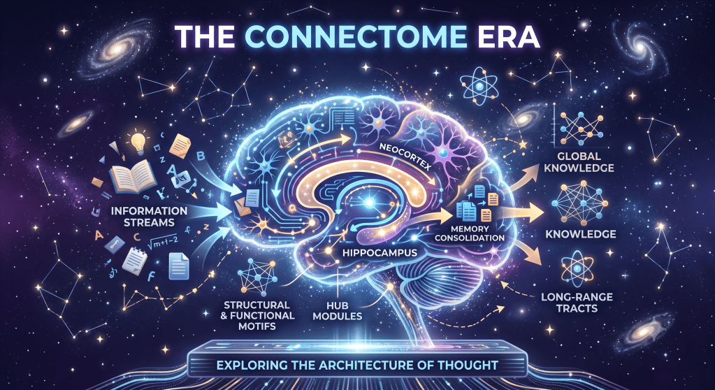
How the Human Connectome Project reshaped neuroscience, revealing the brain as a cooperating network of hub regions rather than isolated functional areas.
In the summer of 2014, neuroscience was in the middle of an identity crisis it hadn’t quite admitted to yet. For more than a century, brain science had operated on a simple premise: find the region responsible for a given function, and you understand the function. Broca’s area handled speech production. The hippocampus handled memory. The amygdala handled fear. This was an extension of the localization tradition — far more scientifically grounded than phrenology, but still focused on assigning functions to individual regions. It had produced real discoveries. But it was also starting to buckle under its own success. The more precisely scientists mapped individual regions, the more obvious it became that no region worked alone. A stroke in one small area could unravel abilities scattered across the brain. A skill thought to belong to a single lobe turned out to depend on a dozen distant partners working in sync. The brain, it turned out, was not a collection of specialized offices. It was closer to a city’s transit system, and cities don’t make sense department by department. They make sense as networks.
This was the atmosphere into which the Human Connectome Project arrived. Funded by the National Institutes of Health and launched in 2009, with major grants awarded the following year to two research consortia, the project set out to do something that sounds almost absurdly ambitious when stated plainly: map the circuitry of the healthy adult human brain in enough detail to understand how its regions actually talk to one another. It was the first of three “Grand Challenges” in the NIH Blueprint for Neuroscience Research, and its backers were explicit that they weren’t just building a bigger atlas. They were trying to launch an entirely new field. As one of the project’s lead NIH scientists put it at the time, the tools produced by the effort were meant to play a major role in launching connectomics as a field in its own right.
By 2014, the project’s early data releases were already reshaping how researchers thought about the brain, and by 2026 — with more than a decade of follow-on studies, lifespan extensions, and computational advances behind it — the “connectome” framework has become close to the default lens through which brain science understands itself. This piece looks at why that shift happened, what the connectome actually revealed, and why thinking of the brain as a network rather than a collection of parts changed not just laboratory science but the way ordinary people talk about attention, mood, and mental effort.
Why the Brain Needed a New Metaphor
For most of the twentieth century, the dominant working metaphor for the brain was modular. Textbooks carried tidy diagrams: motor cortex here, visual cortex there, language centers over on the left. This wasn’t wrong, exactly. Certain functions genuinely do cluster in identifiable regions, and damage to those regions produces recognizable, sometimes very specific deficits. But the modular picture always had trouble explaining a stubborn set of observations. Patients with damage in one region sometimes lost abilities that, according to the map, belonged somewhere else entirely. Cognitive tasks that seemed simple on paper — recognizing a face, retrieving a memory, shifting attention from one thing to another — consistently lit up scanners in multiple, often widely separated areas at once, not just the “assigned” region.
The tension came to a head as functional MRI matured through the 2000s. Researchers studying what the brain does at rest — when a person isn’t performing any task at all — kept finding coordinated activity between regions that had no obvious anatomical reason to cooperate. This resting-state work eventually crystallized into the discovery of the default mode network, a set of regions that quiet down during focused tasks and become active during mind-wandering and self-referential thought. It was one of the clearest early demonstrations that the brain’s organization was fundamentally relational: what mattered wasn’t just what a region did in isolation, but who it was talking to, and when.
By the time the Human Connectome Project got underway, this shift in thinking already had a name in the broader scientific literature: connectomics, the study of the complete map of neural connections in a nervous system. Much of this work borrowed mathematical tools from graph theory, treating brain regions as “nodes” and their connections as “edges,” which allowed researchers to quantify the architecture of the brain much like engineers analyze transportation or communication networks. The ambition of the HCP was to take that concept, which had mostly existed in small, methodologically inconsistent studies, and apply it at a scale and resolution nobody had attempted before.
Building the Map
The scale of the undertaking is easy to state and hard to fully appreciate. The project eventually collected detailed brain imaging data, along with cognitive, behavioral, and genetic information, from over 1,100 healthy adults, including twin pairs and their siblings, a design deliberately chosen so researchers could start teasing apart how much of an individual’s brain wiring is shaped by genes versus by lived experience. The resulting dataset ran to over 27 petabytes, and in a decision that mattered almost as much as the data itself, the project made all of it freely available to researchers everywhere through an open database. That openness turned the HCP from a single study into a shared piece of infrastructure. Labs with no funding to run their own thousand-person imaging study could still ask new questions of the existing data, and thousands of papers have done exactly that in the years since. That decision arguably amplified the project’s impact as much as the imaging itself, since discoveries could continue long after the original scans were completed.
Two complementary imaging approaches formed the technical backbone. Diffusion imaging, a variant of MRI sensitive to the movement of water molecules along nerve fibers, let researchers trace the physical cabling of the brain — the actual bundles of axons connecting one region to another. Functional MRI, which tracks blood flow as a proxy for neural activity, let them observe which regions tend to activate together over time, whether or not a direct physical connection linked them. The distinction matters. Structural connectivity is the wiring diagram; functional connectivity is more like a record of which offices call each other on the phone, sometimes through several intermediaries. The HCP was among the first efforts to gather both kinds of data from the same large group of people, which made it possible to start asking how physical wiring and functional coordination relate to each other, rather than studying either in isolation.
The Hub-and-Spoke Discovery
One of the most influential findings to emerge from this era of research, developed alongside the HCP and later tested extensively using its data, was the discovery that the brain’s most important connections aren’t spread evenly across the cortex. Instead, a relatively small set of regions — later dubbed the brain’s “rich club” — turn out to be unusually densely interconnected with one another, functioning as a kind of central switching network. Researchers Martijn van den Heuvel and Olaf Sporns, whose foundational 2011 paper on the subject helped define this line of work, showed that these highly connected and highly central hub regions play a key role in global information integration between different parts of the network.
Later work extended this picture, showing that hub connections are structurally distinctive: on average they extend over longer distances than other connections, which makes them metabolically expensive to build and maintain, and researchers have argued that this cost is worth paying because these long-range links let signals move between distant regions in just one or two steps rather than many. The practical importance of this rich-club architecture became clearer as clinical researchers started looking at what happens when it breaks down. Disruption to these hub regions has been linked in multiple studies to a range of neurological and psychiatric conditions, which is part of why van den Heuvel and Sporns described the potential clinical relevance of hubs as being underscored by recent studies showing that disturbances of their structural and functional connectivity profile are linked to neuropathology. This is a correlational body of evidence, not proof that hub disruption directly causes any particular disorder, but the pattern has been consistent enough across conditions to make hub integrity a genuine focus of ongoing research rather than a passing curiosity.
Small Worlds, Efficiently Wired
Alongside the rich-club discovery, researchers also found that the human brain exhibits what network scientists call small-world organization: tightly interconnected local neighborhoods linked by a relatively small number of long-range connections. This arrangement appears to balance efficiency with resilience, allowing information to travel rapidly across the brain without requiring every region to maintain a direct connection to every other region. The concept borrows its name from small-world social networks, where any two strangers turn out to be connected through a surprisingly short chain of acquaintances. Applied to the brain, it means a signal generated in one region can typically reach almost any other region in only a handful of steps, largely thanks to the long-range hub connections described above, even though most of a given region’s direct neighbors remain close by.
From Snapshot to Lifespan
The original Human Connectome Project scanned healthy young adults, which was a deliberate and reasonable starting point — a baseline map before adding complexity. But a map of one age group can only answer so many questions. The project’s later phases, collectively referred to as the Lifespan and Disease connectome studies, extended data collection to children, older adults, and people with various clinical conditions, turning a single snapshot into something closer to a developmental timeline.
This extension mattered because brain networks are not static. A study using this lifespan data found that different aspects of brain network organization mature on very different schedules. One recent analysis of what researchers call the “effective connectome” — a measure of how strongly and directionally one brain region influences another — found that this measure follows an overall inverted U-shape development with an average maturation time at around 9 years of age, notably earlier than the maturation timeline seen in simpler measures of functional connectivity. Findings like this are a useful reminder that “the brain matures” is not a single event; different properties of brain organization reach their peak configuration at different points across childhood, adolescence, and beyond, and researchers are still working out exactly what that staggered timeline means for behavior and cognition at each stage.
The genetic side of the project has also continued to yield results. More recent analyses of HCP twin data have found that genetic factors appear to preferentially influence hub-to-hub connectivity, meaning the rich-club connections discussed above show comparatively higher heritability than more peripheral connections in the network. Other integrative work combining structural, functional, cognitive, and genetic data from the HCP has estimated that genetics accounts for roughly 12 to 14 percent of the variation in both structural and functional connectivity patterns, with genetics standing out as the single most predictive factor among the non-connectivity variables researchers examined. These are moderate effect sizes, not deterministic ones — they indicate that genes shape the broad architecture of brain networks without dictating it outright, leaving substantial room for experience, environment, and individual life history to shape the rest.
Why “Networks, Not Regions” Changed the Public Conversation
It would be easy to treat all of this as a story confined to neuroscience journals, but the shift from a region-based to a network-based understanding of the brain filtered outward into how people talk about their own minds. Consider how casually terms like “wired that way” or “brain fog” get used now, phrases that implicitly assume distributed, systemic explanations rather than single-location ones. When popular science writing explains why multitasking degrades performance, or why chronic stress affects seemingly unrelated things like decision-making and immune function, the underlying logic is almost always connectomic: these effects ripple across a network rather than sitting in one box.
This has been, on balance, a clarifying shift rather than a confusing one. The older, modular picture of the brain lent itself to oversimplified public claims — “left-brained” versus “right-brained” personalities being the most persistent example, a distinction that has little support in actual connectivity data, since most complex cognitive tasks recruit both hemispheres working together rather than one hemisphere working alone. A network view is harder to compress into a slogan, but it’s also more honest about how the brain actually operates, and it has pushed science communicators toward more careful language about correlation, integration, and individual variability rather than tidy one-region explanations.
That said, it’s worth being precise about what the connectome data does and doesn’t support. The HCP has documented that healthy brains show considerable individual variability in their network architecture even while sharing broad organizational principles — general patterns of connectivity are consistent across people, but the fine details of any one person’s network are close to a fingerprint. This variability is a genuine, well-replicated finding. It is not, however, license for the popular claim that connectome differences straightforwardly “explain” personality or intelligence in any simple, deterministic sense. Correlations between connectivity patterns and measures like IQ have been reported, but correlation research of this kind describes an association, not a mechanism, and the causal arrows connecting brain wiring, environment, and behavior remain an active and unsettled area of study.
The Computational Turn
If the first decade of the connectome era was about data collection, the years leading up to 2026 have been increasingly about what can be done with that data once artificial intelligence tools are brought to bear on it. Machine learning methods are now routinely used to detect patterns in connectivity data too subtle or too high-dimensional for traditional statistical approaches to catch efficiently. One example that drew attention from NIH leadership involved AI-assisted analysis that helped reveal meaningful sex differences in the way the brain works, a finding that would have been difficult to extract using older methods.
This computational turn has also produced increasingly sophisticated attempts to combine multiple types of data at once — structural wiring, functional coordination, cognitive test scores, and genetic markers — into unified statistical models rather than analyzing each in isolation. Methods with names like Data Integration via Analysis of Subspaces have been developed specifically to let researchers decompose these different data types into shared and individual components, which is a technical way of asking a fairly intuitive question: how much of what makes your brain network distinctive is shared with everyone else, how much is shared with people who resemble you genetically or cognitively, and how much is genuinely yours alone.
Alongside this, the broader field of connectomics has continued expanding beyond the human macroscale connections the HCP focused on. Newer initiatives are attempting to map brain wiring at the level of individual neurons rather than broad regional bundles — an effort sometimes called the “wiring diagram” approach, distinct from the macroscale connectivity maps the HCP specialized in. Projects in this space, including NIH-funded efforts aiming to build complete neuron-level wiring diagrams in animal models, represent connectomics pushing toward finer and finer resolution, even as HCP-style projects continue refining the bigger picture at the scale of whole brain systems.
What the Connectome Did Not Settle
Good science writing about a field this active has to resist the temptation to declare victory. The connectome framework reshaped how neuroscientists think about brain organization, but it did not resolve several deep and still-open questions. Structural and functional connectivity, despite being measured in the same people using the same imaging sessions, do not map onto each other in a simple one-to-one way; two regions can be functionally synchronized without a direct anatomical connection, communicating instead through intermediaries, and researchers are still working out the general rules governing when and how that happens. The relationship between connectome architecture and specific psychiatric or neurological conditions remains largely correlational rather than causal, and translating connectome research into actual clinical tools — diagnostic markers, treatment targets — has proven considerably harder than mapping the connections themselves. And the heritability estimates discussed earlier, while informative, describe population-level patterns; they say very little about what shapes any single individual’s particular brain network, since gene-environment interactions across a lifetime are notoriously difficult to disentangle from genetic contribution alone.
None of this diminishes what the project accomplished. It simply reflects the ordinary trajectory of a genuinely new scientific framework: an initial period of rapid, foundational discovery, followed by a longer, messier period of refinement, contradiction, and slow consolidation. The connectome era is still very much in that second phase.
Conclusion: A Different Kind of Map
What the Human Connectome Project ultimately offered wasn’t a finished picture of the brain so much as a new set of tools and a new default assumption for asking questions about it. Before this era, a natural first question about any cognitive ability was “where in the brain does this happen?” After it, the more natural question became “which network handles this, and how do its parts coordinate?” That’s a substantial shift in framing, and it has proven durable enough to shape not just academic neuroscience but the more casual, everyday vocabulary people use to describe attention, mood, and mental effort.
Looking forward, the trajectory seems likely to continue in two directions at once: outward, as lifespan and clinical connectome studies extend the original healthy-adult map across development, aging, and disease; and inward, as neuron-level connectomics projects push toward a resolution far finer than anything the original HCP attempted. Somewhere between those two scales — the whole-brain network view pioneered in the 2010s and the cellular-level wiring diagrams now under construction — future researchers will likely find the connections that finally let structural maps and functional behavior meet in the middle. That work is unfinished, and probably will be for decades. We still don’t possess a complete map of the mind, but thanks to the connectome era, we finally know that understanding the brain means understanding its conversations, not merely its parts.
Scientific Reference
- National Institutes of Health. “NIH Launches the Human Connectome Project to Unravel the Brain’s Connections.” NIH News Release, 2009.
- National Institute of Mental Health. “Human Connectome Project (HCP).” NIMH Research Initiatives.
- Van den Heuvel, M.P., & Sporns, O. “Rich-Club Organization of the Human Connectome.” Journal of Neuroscience, 31(44), 15775–15786, 2011.
- Watts, D.J., & Strogatz, S.H. “Collective Dynamics of ‘Small-World’ Networks.” Nature, 393, 440–442, 1998.
- Sporns, O. Networks of the Brain. MIT Press, 2011.
- Oldham, S., & Fornito, A. “The Development of Brain Network Hubs.” Developmental Cognitive Neuroscience, 36, 100607, 2019.
- “The Effective Connectome Over a Century of Human Life.” PMC, 2025.
- Bertagnolli, M.M. “Human Connectome Project.” NIH Director’s Blog, 2024.
- Ackerman, et al. “Data Integration via Analysis of Subspaces (DIVAS).” Referenced in Human Connectome Project Overview, EmergentMind, 2024.
- Biology Insights. “The Human Connectome Project: Mapping the Brain’s Network,” 2025.